Mallet Finger | Advanced Orthopaedic Specialists | Fayetteville, AR | Rogers, AR
Advanced Orthopaedic Specialists

January 30, 2018

| AOSHogDocs

Mallet Finger

What is a mallet finger?

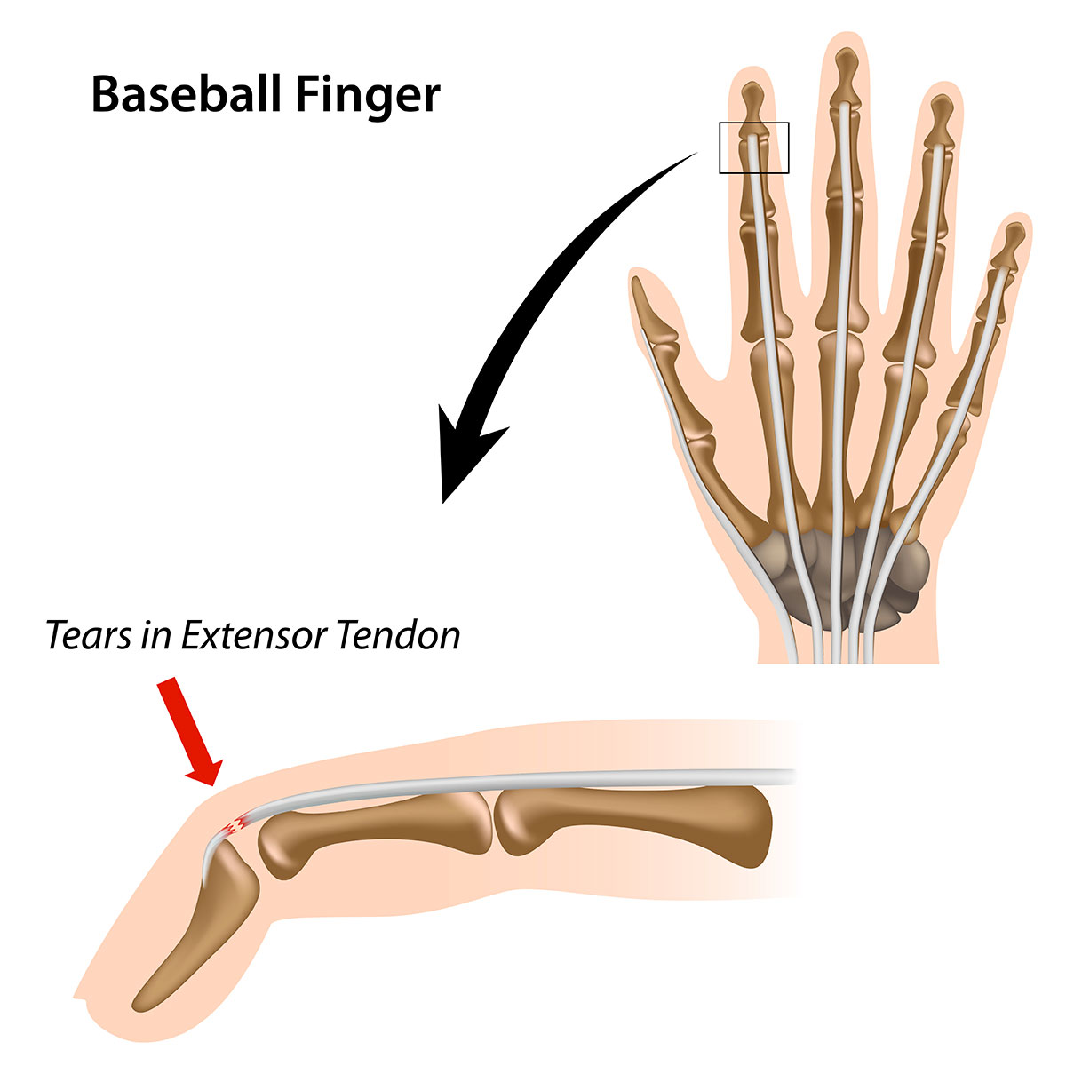
Mallet finger is an injury to the tendon that straightens the last (distal) joint of the finger or thumb. In an extensor tendon injury affecting that joint, the tendon is either torn from its attachment to the last bone or avulsed with a bone fragment from its attachment site. Either way, the anchor is no longer connected to the bone that it needs to move. The result is a finger that lacks strength to fully extend, and stays flexed at rest.

mallet finger

Mallet finger usually occurs from a forced flexion injury to that joint, either with a ball hitting that distal bone (basketball, football, baseball, etc) or with direct trauma, such as a baseball player diving into the bag.

What should I do for a mallet finger?

Treatment is rarely operative, with a large majority of mallet fingers treated non-operatively. The non-operative treatment involves protection and immobilization of that joint in a full-extension splint for 6-8 weeks. That allows the tendon to heal and preserve the function and cosmetic aspect of that distal joint. If there is a large avulsion fracture fragment, or significant subluxation and instability to the distal joint, then operative intervention is warranted.

When should I see a doctor?

Evaluation in the clinic involves examining the affected joint that clinically can make the diagnosis of a mallet finger, and imaging to evaluate for an avulsion fracture. If you are concerned you may have sustained a mallet finger injury, come see us at Advanced Orthopaedic Specialists, and we will get you back on the road to recovery.

March 28, 2017

| AOSHogDocs

Little League Elbow

What is it?

“Little League Elbow” is a disorder of the inner portion of the elbow that affects young athletes.

In this condition the growth plate is irritated and may even separate from the bone. It is caused by repetitive stress on the elbow from throwing too many pitches or possibly using improper mechanics while throwing. The condition is more common in younger athletes who throw curve balls. Other risk factors include throwing more than 80 pitches per game, competitive pitching 8 months or more per year, and continuing to throw despite arm pain and fatigue. This condition is not limited to pitchers, it may also be seen in catchers, infielders, and outfielders.

What are the Symptoms?

Young athletes may complain of pain in their throwing arm. The pain is typically located on the inside portion of the elbow. Athletes may experience more severe pain during the cocking phase of throwing. May also notice decreased throwing speed, accuracy, and distance.

When Should I See My Doctor?

If your child has pain which lasts for several days that it not relieved with rest. Also if you or your child’s coach notice a change in their throwing.

How Can I Prevent Little League Elbow?

It is important to have proper technique while throwing. Throwing curve balls or breaking balls should be discouraged before the age of 14. Coaches and parents should ensure proper rest between outings. Athletes who are pitching in more than one league should be watched cautiously. Parents and coaches should make sure that the athlete is adhering to pitch counts which are appropriate to age.

When Can I Return to Play?

Patients often require 4-6 weeks of rest and rehabilitation. Once pain free the athlete should begin a graduated throwing program before returning to sports.

January 10, 2017

| AOSHogDocs

Lateral Hip Pain – Trochanteric Bursitis

What is Trochanteric Bursitis?

Trochanteric bursitis (also known as greater trochanteric bursitis or GTB) refers to pain at the lateral and posterior aspect of the hip due to inflammation of the bursa, a small fluid filled sac.  This pain usually ranges from mild to moderate, but it can become severe and debilitating at times.  The bursa becomes irritated and inflamed as a result of injury to the neighboring muscles and tendons, or directly from being hit. The iliotibial band (IT band) runs over the trochanteric bursa and is often very tight in people who have trochanteric bursitis.

When the IT band is tight it rubs over the bursa and causes it to become inflamed.  This commonly occurs in active people who run, spin, or lift weights and in football, soccer, cycling, and tennis.  Another contributor to causing trochanteric bursitis is injury to the gluteus muscles as they attach to the bone right near this bursa.  This is often an overuse injury caused by repetitive squatting and/or lunging.  People usually notice a nagging pain on the side of their hip throughout the day or at nighttime in bed when they lay on their side. Typically, the pain is intermittent and only hurts when the area is touched or the muscle causing the issue is used.

How Can I Prevent Trochanteric Bursitis?

Trochanteric bursitis is a result of injury to the surrounding muscles and tendons, so to prevent it you must take care of yourself.  Days of rest are crucial for recover and always important to prevent overuse injuries.  It is recommended to have a least 2 days of rest per week depending on your exercise routine. Often injury occurs because the muscles and tendons are too tight.  The only way to prevent this is to stretch! Remember that cold muscles shouldn’t be stretched and you should always do a slight warm up before stretching.  It’s also important to stretch after exercise as muscles are repetitively contracting during exercise.  The IT band can be difficult to stretch, so a foam roller may be used to help stretch out the IT band.

When Should I See My Doctor?

It’s important to know when to go to the doctor versus treating something on your own. For minor pain, it’s usually a good idea to start with rest and ice for treatment.  If the pain resolves, then you can resume your activity slowly so you do not reinjure the same area. However, if the pain is moderate, reoccurs, or doesn’t improve with basic rest and ice, then it should be evaluated by your doctor.  As always, prompt evaluation and management are the keys to a quick and successful recovery!

July 10, 2017

| Terry Sites, MD

Lateral Hip Pain

Lateral hip pain is common in both active and non-active individuals. This pain tends to be localized over the bony prominence (bump) on the outside(lateral) aspect of the hip. This area will frequently be tender to touch and there may or may not be swelling. The pain may be sharp or dull. The pain may be localized or radiate (travel) down the lateral side of the thigh toward the knee. It may be painful to sleep lying on that side. Most frequently the pain will begin without any known reason.

lateral hip pain

There are several muscles which may be involved with lateral hip pain. The IT band is a muscle which runs from the pelvis to the knee and crosses over the bony bump (named the greater trochanter).When the muscle rubs over the greater trochanter (GT), irritation can occur resulting in a condition called Greater Trochanteric Bursitis. Loss of flexibility in the IT band muscle or overuse can cause this condition. Another muscle that may be involved with lateral hip pain is the gluteus medius (butt muscle). It attaches from the pelvis directly to the GT and this attachment area can become Inflamed, partially torn, or completely torn similarly to the rotator cuff tendon in the shoulder. When this occurs there may be pain or weakness in abduction (spreading the legs). Other conditions which may cause pain about the hip are hip arthritis (usually more in the front or behind), stress fracture, referred pain from low back or lumbar spine, and other pinched nerves.

Evaluation by the specialist will include physical examination and x-rays (to check for arthritis or stress fractures). Treatment for lateral hip pain usually starts with activity modification, stretching exercises and/or oral anti-inflammatories. In many cases the option of cortisone injection with or without physical therapy may lead to quicker relief. In cases that fail to respond to treatment or have been present for a long time an MRI may be considered. Most patients can be successfully treated without surgery. We will work together so you may choose the course of treatment with which you feel most comfortable. You and I can chase that hip pain away!!!

December 10, 2018

| Christopher Arnold, MD

I hear about “stem cells.” Do they work? Can it save my knee?

The promise of stem cells isn’t necessarily new. Academics have been studying them for more than 20 years. But starting in the last decade, cell therapy pioneers have taken them out of the lab and begun to use them for various health conditions. Accumulating data suggests that stem cell injections are an effective way to treat joint pain and injury including arthritic knees, torn ACLs and rotator cuffs, and more.

As you hear more about this type of non-surgical treatment, you may have a growing list of questions. It’s good to be informed, and as a practice that has carefully studied and has performs stem cell injections on a regular basis, we want to help answer some of the most common questions.

What exactly are stem cells?

Stem cells are cells that are capable of differentiating or changing into other cells. In the knee, the hope is that these cells will develop into cartilage and improve the arthritic knee.

Are there different types of stem cells?

Yes, stem cells are currently available in the United States from three different sources, and we’d like to be clear on each:

1. Amniotic (from the placenta)

  • Commonly used by providers not familiar with cell therapy
  • Are not FDA approved for joint injections
  • Are not living cells
  • There are no studies supporting these for joint injuries
  • Advanced Orthopaedic Specialists does not use these

2. Adipose (from fat)

  • Have a high concentration of stem cells
  • Are not FDA approved for joint injections
  • Advanced Orthopaedic Specialists does not use these

3. Bone marrow aspirate

  • These have living stem cells in addition to a large concentration of growth factors
  • They are FDA approved
  • There are multiple studies supporting their use in joints
  • Advanced Orthopaedic Specialists offers these

stem-cells-knee-blog-post-supplement

Will stem cells grow new cartilage?

Studies show that stem cells won’t restore the cartilage to normal, nor make an arthritic knee normal. However, studies do show they can slow down the progression of arthritis and make the remaining cartilage less likely to deteriorate.

If stem cells don’t grow new cartilage, then how do they work?

Bone marrow aspirate has not only stem cells, but also a variety of growth factors that create a healthier joint. In every joint there are two types of growth factors. Some that build it up (anabolic growth factors) and some that tear it down (catabolic growth factors). A normal joint has a high concentration of anabolic, and a low concentration of catabolic growth factors. An arthritic joint has the opposite (high catabolic and low anabolic). Once a joint experiences injury, the catabolic growth factors become predominate and destruction ensues. When bone marrow aspirate is injected into the joint, the hope is to reverse this destruction and create a healthier joint.

I see so many providers advertising stem cells. How do I know who to choose?

Because this is still a fairly new treatment, it is not covered by insurance. That means patients have to pay cash for this service. Unfortunately, there are some providers out there who are taking advantage of patients for financial gain at their patients’ expense. In addition, they may be using non-FDA approved treatments on patients who are not even candidates for this service. If you elect to have stem cell injections, make sure you have them performed by a physician who uses FDA-approved injections, and confirm that you truly are a candidate before spending the money.

How do I know if I’m a candidate for stem cells?

To be a proper candidate, studies show you must:

  • Not have bone-on-bone contact on your X-ray
  • Not be obese
  • Not smoke
  • Not had any meniscus tears removed
  • Be in general good health


Are these injections better than cortisone?

There are two types of injections. One that helps with the pain, but does not improve the joint (cortisone, Toradol, and viscosupplementation). The other helps with pain, but also makes a healthier joint (PRP and bone marrow aspirate). So, while cortisone can be a great treatment to manage pain, it does not ultimately improve the joint.

What is PRP?

Platelet Rich Plasma (PRP) contains growth factors, but no stem cells. Studies show it can be better than viscosupplementation, but not as good as bone marrow.

How and where do I get a PRP?

The patient has blood drawn from the arm in the office, then the platelets are separated out with a centrifuge, then injected into the joint. Platelets have a high concentration of good growth factors that facilitate healing and decrease inflammation.

How and where do I get a bone marrow aspirate?

An orthopedic surgeon aspirates bone marrow from the patient in the office under local anesthesia (he injects the pelvis with lidocaine). Once the cells are retrieved, they are injected into the joint under ultrasound. The patient takes a pain pill and valium 30 minutes before the procedure. After the procedure they are given crutches for 3-5 days. In one week, they walk normally. No heavy impact activities for 6 weeks.

How do I learn more about this procedure?

Advanced Orthopedic Specialists offers regular, free seminars with our board-certified surgeons to provide you with medically accurate information and to help you understand your options. Sign up to get notified about our next session.

If you’d like a private consultation with one of our specialists, schedule an appointment by calling us at 479-966-4187 or contact us online.

Written by Dr. Christopher Arnold

February 14, 2017

| AOSHogDocs

Knee Osteoarthritis

What is Knee Osteoarthritis?

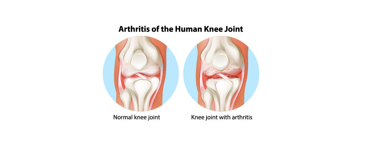
Knee osteoarthritis (OA) is the wear and tear type of arthritis that develops over the years. It tends to be accelerated in anyone who has had an ACL tear in the past or other significant trauma to the knee. OA first starts as softening of the bone cartilage cap and then it continues to erode down to bare bone. It can be very painful at times or silent. As pressure is put on the areas of arthritis, it becomes inflamed and causes pain and swelling in the knee. Patients commonly describe the pain as a dull ache or a sharp pain in the front/inner portion of the knee. Due to the inflammation people often feel their knee is stiff, especially in the colder weather. Another common finding with OA is feeling crepitus. Crepitus is the “creeks and cracks” that people feel in their knee when walking or climbing stairs. OA is progressive and will continue to worsen, however the rate at which OA worsens varies amongst individuals.

knee osteoarthritis

How Can I Prevent Knee Osteoarthritis?

Knee osteoarthritis is very difficult to prevent, but there are certain variables that can be change to help. The first modifiable risk factor is someone’s weight. For every pound that a person loses it decreases about five pounds of force through the knee. For example, losing 10 pounds will decrease about 50 pounds of pressure through the knee. This force reduction can make a tremendous difference. The force transmitted through the knee can also be decreased by choosing activities that are easier on the knees. Instead of running on the road consider a softer surface, biking, or swimming. If a person has a gym membership, then consider using the elliptical machine. Shoes also play a large role in foot strike pattern and how force is transmitted through the body. Always make sure your shoes are not worn out and fitted properly to your foot and walking pattern. Finally, some studies have shown that glucosamine and chondroitin supplements may help preserve cartilage. Not all the medical literature has demonstrated this to be true, but some people tend to find it helpful.

When Should I See My Doctor?

Knee OA is progressive and it’s important to have it under control, so you can enjoy your life. If the knee is swelling, then you should instinctively use ice. Ice will help control the inflammation and the subsequent pain that develops. Over the counter anti-inflammatories may also be help to again control pain and inflammation. If the pain persists despite these basic treatments, then you should see your doctor. Other types of treatments can be provided to help osteoarthritis. Injections can be used to reduce pain and inflammation or to restore cartilage and reverse arthritis. As arthritis becomes severe then options become limited and a joint replacement may be indicated. Don’t be a victim of knee osteoarthritis and its pain, please see a doctor to make sure your arthritis is under control.

April 11, 2017

| Christopher Arnold, MD

I TORE MY TOMMY JOHN, WHAT ARE MY OPTIONS?

The “Tommy John” ligament is technically called the ulnar collateral ligament of the elbow.  It is the primary stabilizer to the elbow in a throwing athlete as the elbow is flexed between 20 degrees and 120 degrees.  It gained its name, the Tommy John Ligament, in 1974 when a professional baseball player Tommy John tore his ulnar collateral ligament and subsequently had it reconstructed, he was able to return to full pitching activities. Ulnar collateral ligament injuries are most commonly seen in baseball players, but they are not unique to this sport.  They also are seen in football, Javelin throwing, volleyball, tennis, gymnastics, wrestling, water polo, and tennis.  An injury to the Tommy John can occur either from continuous overuse or from a single traumatic event.  When a patient tears the Tommy John, there is usually is significant pain on the inside of the elbow with overhead activities.  There are two types of tears.  Most commonly, we see partial tears.  These are seen most likely with repetitive overhead sporting activities.  There also is a complete tear.  This can be seen with repetitive activities, but most commonly occurs after a traumatic incident.  The orthopedic community has seen a dramatic rise in the diagnosis of ulnar collateral ligament injuries over the past 10 years as there has been an increased level of activity in the sports which produce these injuries.  Baseball has become a year-round sport.  There has been an effort to limit the injuries to the elbow by limiting the number of pitches players can make during a game or during the weekend.  Despite this, Tommy John injuries are still very common.

What are my treatment options?

At Advanced Orthopaedic Specialists, there are basically two treatment options for the injured ligament and this hinges on if the ligament is partially torn or completely torn.  If there is a partial tear of the ligament, we recommend nonoperative treatment.  We treat the patient with physical therapy, anti-inflammatories, and a prolonged period of rest.  Once the patient becomes asymptomatic, we will start a graduated throwing program in the hopes of return.  We have had excellent results with partially torn Tommy John ligaments with this regimen.  It also is an option for a partially torn ligament to perform a stem cell injection into the ligament.  We commonly use platelet rich plasma or “PRP.”  This technique is done in the office where a sample of the patient’s blood is drawn.  We centrifuge the blood and remove the platelets and inject these into the injured site under ultrasound guidance.  With a PRP injection into the Tommy John ligament, we typically follow this with a period of rest, immobilization, and then after approximately one month, a gradual return to sports with a graduated throwing program.  We feel strongly that in partial tears less than 50% of the ligament, the athlete can have an excellent result and return to full play with a conservative program.  If a ligament is torn more than 50%, we still attempt this regimen; however, success is not quite as good.

If the patient has complete tear or if they have failed nonoperative treatment for a partial tear, it is an option for surgical reconstruction.  Reconstruction of the ulnar collateral ligament is commonly performed at Advanced Orthopaedic Specialists.  We have performed this in the junior high, high school, and collegiate-level athletes and anticipate a full return to sport.  The ligament is reconstructed with a tendon, which is taken from the forearm of the patient and inserted into the place of the old ulnar collateral ligament.  The surgery takes approximately one hour and the patient is discharged home the same day.  The patient is placed into a splint for one week and then into a brace that allows range of motion.  The brace is discontinued at six weeks.  Physical therapy is started at the one week point.  With physical therapy, we go through a series of steps with passive range of motion where the therapist moves it, followed by active range of motion where the patient moves it, followed by strengthening.  At approximately 14 weeks, we slowly resume a throwing program.  Most athletes, with the exception of pitchers and catchers, are able to return to their sport at approximately six months.  It typically takes a pitcher or catcher approximately 9 to 12 months before they are able to return to full play.  The success with an ulnar collateral ligament reconstruction is approximately 90%, good to excellent, were the patient can return to their level of playing.

If you have been told you had a Tommy John ligament injury, please contact us at Advanced Orthopaedics Specialist where we specialize in the treatment of the injured athlete.

May 17, 2019

| Zac Snow, DPT

Is Exercising Really That Important?

I want to preface this blog post by stating I have a huge bias in favor of exercise. As a physical therapist, I get to use this amazing tool every day to guide others to their most robust state. Yes, it takes some training to know what is safe and what is not for someone that is injured or just had surgery, but for those who are healthy, this can be a lifestyle that pays dividends.

A Little Goes A Long Way

The medical profession has known for years that physical activity, even a little bit, can drastically change a patient’s health risks. About $117 billion in annual health care costs and about 10 percent of premature mortality are associated with inadequate physical activity. Strong evidence demonstrates numerous health benefits for children and adults.

Children under the age of 17 can benefit from improved bone health, weight status, cardiorespiratory and cardiometabolic status, cognition and reduced risk of depression.

Adults and older adults can benefit from physical activity in numerous ways, as well. Physical activity in adults lowers the risk of all-cause mortality, hypertension, type 2 diabetes, cardiovascular disease, cancers, dementia, anxiety, depression, and falls. In addition, physical activity improves sleep quality, bone health, weight management, cognition, and general quality of life.

Your Body and Mind Will Thank You

Improved physical function in older adults reduces the risk of falls and fall-related injuries and contributes to their ability to maintain independence. It is also true for young and middle-aged adults, as improved physical function helps them more easily accomplish the tasks of daily living, such as climbing stairs or carrying groceries.

In addition to improving physical function, physical activity may improve cognitive function among youth, adults and older adults. Aspects of cognitive function that may be improved include memory, attention, executive function (the ability to plan and organize; monitor, inhibit, or facilitate behaviors; initiate tasks; and control emotions), and academic performance among youth.

Recommended Amounts

As of 2018, the United States Department of Health & Human Services (HHS) recommends the following:

  • Children age 3 to 5 years should be active throughout the day with a variety of activities to stimulate cognitive and physical development. 
  • Children age 6 to 17 years should be moderate-to-vigorously active 3 or more days per week for 1 or more hours per day consisting mostly of aerobic activity with some muscle and bone stimulating activity. 
  • Adults should aim to spread out 2.5 to 5 hours of moderate-intensity, or 1.25 to 2.5 hours of vigorous-intensity aerobic exercise per week. Additional health benefits can be gained by greater than 5 hours per week of moderate-intensity aerobic activity, as well as strength training 2 or more days per week.
  • Older adults benefit from the same activity guidelines as their younger counterparts. However, older adults benefit additionally from balance training throughout the week.


Keeping It Simple

Exercise and physical activity can be confusing and intimidating. To simplify things the HHS has laid out guidelines for both exercise intensities and types. By combining the two on a weekly basis a person can easily manage their goals of staying physically active and healthy. Below are examples of age-based exercises that can be paired with the frequency and durations above.

Children age 6 to 17 

  • Moderate aerobic: brisk walk, casual biking, dancing, skipping 
  • Vigorous aerobic: running, basketball, sustained swimming, cycling
  • Strengthening: climbing trees or playground equipment, tug-o-war, structured weight lifting
  • Bone development: any activity (aerobic or strengthening) that produces a force on the bones (i.e. landing a jump, running, weight lifting, jumping rope)

Adults

  • Moderate aerobic: brisk walking, biking less than 10mph, doubles tennis, yoga, water aerobics, Zumba, yard work, dancing
  • Vigorous aerobic: running, lap swimming, cycling, jump rope, hiking uphill, kickboxing, heavy yard work, basketball, high-intensity interval training (HIIT)
  • Strengthening: heavy yard work – lifting, pushing or pulling heavy loads, weight training – resistance exercise of the hips, knees, shoulders, back and core

Older Adults have the same exercise recommendations as their younger counterparts with the caveat that they attenuate the intensity to their current state of health, as well as focusing on balance.

  • Balance activities: Tai Chi, Yoga, Dancing, Tennis, Zumba

Ask Your Physician

You’ve heard it before but always consult a physician prior to starting an exercise regimen. A physician that works with an active population would be ideal, such as a fellowship-trained sports medicine physician (Drs. Ylanan, Wagner, & Balle). They can screen you for prior health risks that may not be apparent to you or a personal trainer.

If you’re interested in exercise and have a health condition (or not), a physical therapist will work with you to determine what activities and exercises are safe and educate you on what you can expect long term.

It’s Never Too Late

If you’re interested in learning more about physical activity guidelines there is a wealth of knowledge in the “Source” link below. Remember, something is better than nothing, and it’s never too late to start!

Sources: Physical Activity Guidelines for Americans 2nd Edition (2018)

Written by: Zac Snow, DPT

June 21, 2017

| Christopher Arnold, MD

I Have Torn My Labrum In My Shoulder, What Should I Do?

The labral tear in the shoulder is a very common injury to our athletes seen at Advanced Orthopaedic Specialists. The shoulder is made of a ball and socket joint. The ball is very large. The socket is very small. The joint is very similar to a golf ball, which rests on the tee. The stability of the ball onto the socket is dependent on the labrum and the ligament which attaches to the labrum. This offers the primary stability to the shoulder joint. Secondary stabilizers include the rotator cuff muscles, which surround this. The socket is very similar to the face of a clock and there is a labrum, which surrounds this completely. There is a superior labrum, which is at the top or the 12 o’clock position. There is the anterior labrum, which is in the front part of the socket and there is a posterior labrum. In the back, the labrum is cartilaginous and has the appearance of a “gristle.” It is very similar to the structure which makes up the tip of the nose. The labrum acts as a buttress to keep the shoulder in joint. More importantly, the ligament which stabilizes the shoulder attaches to the labrum.

There are basically three types of labral injuries, which are seen in the athlete. There is a superior labrum. This is commonly seen with repetitive throwing or repetitive overhead activities. There is an anterior labrum, which also is seen with repetitive throwing and overhead activities, but is also very common with a shoulder dislocation. There also is a posterior labrum, which is in the back part of the shoulder and this is commonly seen in direct force to the shoulder or in heavy weightlifters and offensive lineman.

In our patients who tear their labrums, initially, a nonoperative protocol is started. We start with trying to strengthen the surrounding structures, i.e., the rotator cuff. The patient is started on anti-inflammatories and possibly a corticosteroid injection. Once the patient has resumed their full range of motion and has regained their normal strength, we allow the patient to return to play. If the patient experiences persistent pain or instability, we can consider surgical options. Surgery for the superior labrum consists of either a simple debridement. At which point, a microscope was inserted in the shoulder and the cartilage tear is cleaned up. This is typically a very quick return to sport, as soon as three months. If, however, the superior labrum is completely torn, we attempt to repair this and return to sport is approximately four to six months.

If an anterior labrum is torn, again, we try to treat this nonoperatively with physical therapy, anti-inflammatories, and possibly injection. If, however, the patient continues to be symptomatic or if they experience instability, excellent results have been obtained with labral repairs.

In the past, this procedure was done through a large incision. We currently do this through the microscope where we can reattach the labrum to the normal sockets. The procedure takes approximately one hour. Physical therapy is started immediately. A sling is worn for approximately four to six weeks. A strengthening program is started at three months and return to sport is typically four to six months. Anterior labral repairs have approximately a 90% to 95% success rate.

A posterior labral tear also is treated initially nonoperatively; however, if the patient continues to be symptomatic, we perform this in a very similar fashion to the anterior labrum with a similar return to sport timetable.

With the advent of arthroscopy, the labrum is easily repaired and we can anticipate a much better result as opposed to previously when was done via an open fashion. At Advanced Orthopaedic Specialist, we have repaired hundreds of labrums and have allowed our patient to return to full level activity in a predictable fashion. If you have been told you have a torn labrum, please contact us at Advanced Orthopaedic Specialists.
October 18, 2017

| Christopher Arnold, MD

I have arthritis in my knee. What are my options?

Arthritis of the knee is a condition which is commonly suffered by millions Americans each year. Technically what arthritis is, is any inflammation or damage to the knee cartilage. There are two types of cartilage in the knee. The meniscus which is the main cushion between the femur and tibia. The second type of cartilage is articular cartilage. Any damage to the articular cartilage is technically considered “arthritis”. There are various causes of arthritis. Most commonly is wear and tear or osteoarthritis. However, also we can see a traumatic arthritis, or an inflammatory arthritis and condition such as rheumatoid arthritis, gout and other similar entities. Damage to the articular cartilage is typically a progressive disorder and the end result is loss of all of the cartilage with bone-on-bone contact. The patient with bone-on-bone contact experiences significant pain about the knee joint, itself and is typically described as a dull toothache, constant pain. Advanced Orthopaedic Specialists (AOS) treats thousands of patients each year with arthritis of the knee. We are commonly asked what the different options are. There is essentially two options. Nonoperative and operative. Before moving forward with operative treatment, we focus on the nonoperative treatment of arthritis. Typically, we recommend activity modifications, avoiding heavy impact activities, and a weight loss program. If this fails, we place patients on medications such as anti-inflammatories or Tylenol. We avoid narcotics in patients that have arthritis in the knee. A brace is a common option for the arthritic knee as it can help to stabilize the knee and to take pressure off of the area of arthritis.

Should the aforementioned fail, we have had excellent success with various types of injections. Corticosteroid injections or (cortisone) are very safe and easy to do. These are safe to do every three months for the patient. They have very little associated risks and can help to get rid of the inflammation and subsequent pain within the knee. These are safe as long as given no sooner than every 3 months and as long as they are not continuing to be administered while they are no longer working. Another injection to improve the inflammation is a “Toradol injection.” This is similar to cortisone but has more of an anti-inflammatory effect and again, can be given every 3 months.

Viscosupplementation is very commonly used for arthritic knees that don’t have “bone-on-bone contact.” These are typically approved by the insurance companies. It’s a series of 3-5 injections. Its purpose is to lubricate the knee, to stimulate the knee to make more of a normal lubricating fluid and also has a pain-relieving or anti-inflammatory effect. These typically work well for arthritis that is non-end stage.

A more recent type of injection at AOS are stem cells. There are a variety of stem cells as outlined below:

A. Platelet Rich Plasma. This is where blood is drawn from the patient and the platelets are isolated and injected back into the knee. We have had excellent success with this. It is an in-office procedure.
B. Amniotic stem cells. This is an off the shelf stem cell injection, again with excellent results.
C. Adipose stem cells injection. This is a procedure done in the office where some stem cells are taken from adipose or “fat tissue” from the patient. The stem cells are then spun down within the office and injected into the knee joint.
D. Bone marrow aspirate stem cells. This is done in the office as well with local anesthetic where some bone marrow cells are aspirated from the patient’s pelvis and the stem cells are isolated and injected into the knee. We have had excellent results with all four of these stem cell modalities. Unfortunately, these are not covered by insurance.

If all of the aforementioned fails and the patient continues to have significant knee pain, the next step would be a total knee arthroplasty. Advanced Orthopaedic Specialists does over 500 knee replacements per year. If arthritis isolated to one of the three portions of the knee, the patient may be a candidate for a partial knee replacement or a unicompartmental arthroplasty. If the arthritis is located to two of the three compartments, then a total knee is necessary.

Technology has advanced dramatically in the modalities of total knee arthroplasties. They typically take 1 hour to perform. Some patients are able to go home the same day whereas the majority go home the following day or two days later. The infection rate for total knees across the country is 1-3%. AOS has an infection rate of approximately 0.5%. We feel that this is because of the multitude of total knees which we perform and the system which we use to perform the total knee replacements. A total knee is performed in approximately one hour and although the patients thinks that the doctor cuts off the entire end of the thigh in the shin, it actually is more of a “resurfacing arthroplasty.” We do it through a less invasive incision and place a cap on the end of the femur as well as on the tibia and behind the patella with plastic in between. The patient walks on the knee approximately one hour after the surgery and is discharged to home once they are independent in therapy which ranges from the same day to two days later.

If you have disabling knee pain, please contact Advanced Orthopaedic Specialists to learn of your treatment options.