Your Complete Guide to Fall Prevention and Action | Advanced Orthopaedic Specialists | Fayetteville, AR | Rogers, AR
Advanced Orthopaedic Specialists

September 23, 2019

| Zac Snow, DPT

Your Complete Guide to Fall Prevention and Action

With fall just around the corner, it’s also time to start thinking about falls. As an interesting fact, September 23rd is the first official day of fall in the Northern Hemisphere, as well as Fall Prevention Awareness Day. For adults 65 years or older, falls are the leading cause of fatal and non-fatal injuries leading to broken hips, wrist fractures, head injuries and decreased quality of life. Indirectly, falls and fear of falling can lead to decreased social engagement, declining independence, and poor mobility thereby affecting mental health and contributing to feelings of helplessness.

A Look at the Statistics

Annually, 1 in 4 older adults will experience a fall resulting in approximately $50 billion dollars spent treating the aftermath. Most of these costs are hospital-related with over 800,000 older adults being admitted annually at a rate of 1 every 11 seconds with fatalities resulting once every 19 minutes. The financial cost of these traumas, approximately 75%, falls on Medicare and Medicaid. If falls rates are not reduced, direct treatment costs are projected to reach $101 billion by 2030.

Risk Factors to Consider

Falling is not a normal part of aging, but as people get older a multitude of factors contribute to injuries or death as a result of a fall. Below are risk factors. The more you have, the greater the risk of falling.

  • History of falls
  • Being female
  • Sedentary lifestyle
  • Overall poor health
  • Recent hospitalization
  • Advanced age
  • Poor balance, due medical condition, such as stroke, Parkinson’s Disease, Multiple Sclerosis, diabetes
  • Leg weakness
  • Easily fatigued
  • Limited flexibility
  • Problems with vision or touch sensation (diabetic retinopathy or peripheral neuropathy)
  • Getting dizzy when you stand up from sitting or lying down (orthostatic hypotension)
  • Trouble with memory and thinking
  • Joint pain/arthritis
  • Taking too many medications or taking certain types of medications
  • Depression and/or anxiety
  • Home hazards, such as throw rugs, poor lighting, or a lack of handrails on stairs
  • Inappropriate footwear
  • Inappropriate use of a walker or a cane


Can a Physical Therapist Help?

Yes! Many risk factors for falls are modifiable, and physical therapists are specifically trained to recognize and address complicating factors that directly and indirectly contribute to falls. Even during routine evaluations, unrelated to falls, a physical therapist may ask older adults about their fall history. I always tell my patients it’s much easier to address balance than to recover from a hip surgery.

How Does a Physical Therapist Assess and Treat Balance and Falls?

First, we gather your current and previous history. A physical therapist will start with questions regarding fall history, activity level, medications, sensations of dizziness, faintness when changing position or feelings of fatigue with ambulation. In addition to physical symptoms, your physical therapist will address your environment, such as footwear, assistance at home, and home layout. We look to you to help identify what contributing factors you’re already experiencing.

Once we have your history, we move onto your physical presentation. For this portion we exam lower body strength, flexibility, sensation of the feet (specifically for diabetics), and vision. Most importantly, we assess your balance through formal and informal testing. During a formal balance assessment, a physical therapist will utilize a test, such as the Berg or Tinetti to generate a score. This score is taken upon initial evaluation and periodically retested to assess your improvement as well as determine the level of fall risk. Informal testing consists of gait analysis, observation of transfers, and lower body structure.

Following the initial evaluation, your physical therapist will recommend what you can safely begin at home. We will develop a formal plan of care specific to your needs that will be carried out in the clinic. In addition, you will also receive a home exercise program as well as recommendations to make your home environment safer and easier to navigate.

Six Exercises to Help Prevent Falls

These exercises help prevent falls by challenging your balance in a safe manner. If you’re unable to perform these exercises then you may want to have your balance formally assessed.

  1. Standing March: Stand in place and start marching in place slowly for 20-30 seconds. As this becomes easier, challenge your balance and change up the surface you are marching on: from hardwood to carpet, foam pad, grass, etc.
  2. Standing 3-Way Kicks: Standing on 1 leg (with a soft knee, not locked in full extension), move the other leg in front of you (keeping your leg as straight as possible), then out to the side, and then behind your body. Perform 10 times on each side.
  3. Sidestepping: Walking alongside (facing) a countertop or near a wall (with hands on the surface as needed), step sideways in 1 direction with your toes pointed straight ahead. Move 10 steps in 1 direction, then return in the other direction. As this becomes easier, use a resistance band just above the ankles.
  4. 1-Leg Stand: Stand on 1 leg as long as you are able, up to 30 seconds. Alternate legs, and try to do this 3-5 times on each leg. As this becomes easier, challenge yourself by doing other tasks while standing on 1 leg, such as brushing your teeth, talking on the phone, or while doing biceps curls.
  5. Sit to Stand: Rise out of a chair without using your arms to push up. If this is difficult at first, use a firm pad underneath you (to place on chair seat) to raise you as you need. Perform 10 times.
  6. Tandem standing or tandem walking: Place 1 foot directly in front of the other, so the heel of the front foot touches the toe of the back foot. Maintain standing in this position as long as you are able, or up to 30 seconds. As this becomes easier, try taking a few steps in this heel-to-toe format, as if you are walking on a tightrope. Remember to use something to hold on to for safety.

With falls being a significant cause of morbidity for older adults, there is an abundance of resources for patients, family members, caregivers, and healthcare professionals. Listed below are various resources based on the Stop Elderly Accidents, Death & Injuries (STEADI) Initiative from the Centers for Disease Control and Prevention (CDC) and the University of North Carolina.

Resources For Patients

STEADI Initiative / Postural Hypotension
Stay Independent Brochure
What You Can Do to Prevent Falls  

RESOURCES For Caregivers/Family Members

STEADI Initiative / Protecting Loved Ones
Check for Safety

RESOURCES For Healthcare Professionals


STEADI Initiative / Evaluation Guide for Older Adult Clinical Fall Prevention Programs


Source Material

Falls Prevention Awareness Day 2018 Impact Report. (2018).
Mulcahy, J. A., PT, MPT. (n.d.). 6 Balance Exercises You Can Safely Perform at Home.
Shubart, T., PT, PhD. (2011, June 2). Physical Therapist’s Guide to Falls.

February 10, 2020

| Zac Snow, DPT

What You Need to Know About Low-Impact Activities

Low-impact activities are defined as any type of exercise which promotes physical fitness but does not stress the musculoskeletal tissues, bones or joints. Conversely, high-impact activities are the opposite in which they place a significant strain on the musculoskeletal system due to their high velocity and ballistic nature. Low-impact activities can be thought of as “joint sparing” since they apply little to no strain on the bones and joints of the body. These activities can be further broken down into low-impact exercise and low-impact sports.

Low-Impact Exercises

Exercise is an essential element of leading a healthy lifestyle. There are many options to explore when looking for low-impact options. The most accessible form of low-impact exercise is simply walking. This can be done indoors or outdoors, day or night, fast or slow, uphill or downhill. It is easily manipulated to increase the time, distance, or pace to further benefit the person. Other forms of low-impact exercise include: 

  • Tai Chi
  • Yoga
  • Pilates
  • Swimming
  • Aqua aerobics
  • Hiking
  • Row machine
  • Indoor cycling
  • Strength training: weights, bands, body weight
  • Elliptical
  • StairMaster®


Low-Impact Sports

In addition to low-impact exercise, there are low-impact sports. These can be either individual or team sports. These will not target specific body areas like exercise will, but they are an excellent way to remain active while minimizing the risk of joint damage. These exercises include:

  • Swimming
  • Horseback riding
  • Cycling
  • Rowing
  • Cross country skiing
  • Water polo
  • Kayaking
  • Golf
  • Ballroom dancing


Benefits of Low-Impact Activities

There are numerous benefits of low-impact activities with the most obvious being that there is a relatively low risk of bone, joint or muscle injury. Due to the fact that low-impact activities place a less intense demand on the body, they can typically be sustained for long durations or performed daily without risk of injury. There are also certain populations where low-impact activities are preferred. Individuals diagnosed with osteoporosis, osteopenia, osteoarthritis, and women who are pregnant.

Low-impact activities can come in the form of open-chain or closed-chain activities. That is to say that the extremity being used is free to move or is fixed, respectively. A squat would be an example of a closed-chain exercise while a seated knee extension would be an example of an open-chain exercise. Those diagnosed with osteoporosis and osteopenia benefit most from closed-chain, low-impact activities due to the bone-stimulating effect induced by weight-bearing. Those diagnosed with osteoarthritis will also benefit from closed-chain, low-impact activity. The caveat being that the load and/or range of motion needs to be monitored closely to prevent initiating a painful inflammatory process in the joint.

PHYSICAL THERAPY Experts

Low-impact activities are one of the many modes of treatment utilized in physical therapy. This mode of exercise allows for loading of painful, sensitive tissues without the risk of injury or increasing pain. If you are unsure of how to properly execute a low-impact exercise regimen or if a low-impact sport is safe for you, a physical therapist can help. Physical therapists are experts in human movement and the effect exercise has on the body while accounting for age, gender, and medical history. Advanced Orthopaedic Specialists has a staff of highly trained physical therapists and would love to help you get moving again. Schedule an appointment today!

April 10, 2018

| AOSHogDocs

What type of Ankle Support or Brace is best for your injury?

Commonly when you have injured your ankle, your treating physician will place you into an ankle brace or support to help while you are recovering from your injury.

So what’s the difference between a brace and a support, and when should each one be used?

Ankle Brace: This type of Durable Medical Equipment piece will usually be prescribed for ligament injuries or with fracture care along with some tendon injuries. They will usually have straps and laces to limit certain motions of the ankle. These types of braces provide the support to protect the ligament(s) or the healing fracture.

Ankle Support: This type of Durable Medical Equipment piece will generally be prescribed for milder injuries to the ankle that could include injuries such as tendinitis or bursitis. These are usually a wrap or sleeve that gives compression and warmth as well as giving a form of biofeedback to the patient.

No matter what type of brace or support your Physician prescribes you should use it as another tool in the recovery process of your injury.

November 18, 2019

| Natalie Jobe, APRN

What’s The Best Nutrition Before and After Surgery?

A healthy and well-balanced diet is essential not only for everyday living but also in preparing and recovery from surgery. Having a well-balanced diet before and after surgery helps you to have a speedier recovery and decreases your chances of infection and wound healing difficulties. Some essential nutrients to have in your pre and post-surgical diets include; fiber, protein, carbohydrates, fat, vitamins and minerals and proper hydration.

Fiber

A common complaint post-surgery is constipation. This is caused by immobility and pain medication. To help alleviate this issue it is important to consume high-fiber foods before and after surgery. Foods high in fiber include fresh fruit and vegetables, whole grains and oatmeal. Avoid processed foods, cheese, and dairy products. Staying hydrated by drinking plenty of water post-operatively will help alleviate this as well.

Protein

Protein is essential for energy to help you recover and heal from your surgery. Lean meats such as chicken, turkey, pork and seafood are all excellent protein sources post-surgery. These protein sources have amino acids that are vital for adequate wound healing and tissue regeneration.

Carbohydrates

Post-surgery many patients experience fatigue from the procedure. This can be counteracted with helping carbohydrates to boost your energy. Good sources of healthy carbohydrates are whole grains, fruits, vegetables, beans, and legumes. Avoid processed bread and sweets.

Fat

Healthy fats can improve your body’s immune response and speed up healing and decrease your risk of infection. Healthy fats are found in olive oil, coconut oil, avocados, nuts, and seeds.

Vitamins and Minerals

Vitamins and minerals are essential to a healthy and quick recovery after surgery. Vitamin A and C are important for wound healing. You can find these in oranges, berries, potatoes, dark green leafy vegetables, carrots, melons, kale, spinach, tomatoes, and bell peppers. Vitamin D is essential for bone health and can be found in milk, fish, eggs, and fortified cereals. Zinc and Iron are both important for wound healing and energy post-surgery. Zinc can be found in meat, seafood, dairy, and beans. Iron is found in meat and poultry, apricots, beans, eggs, and whole grains.

Hydration

Staying hydrated after surgery is important for your recovery as well. You can stay hydrated by drinking at least 8 glasses of water every day after surgery. Adequate hydration helps prevent constipation and helps aid in the absorption of medications and vitamins/minerals needed for recovery.

If you have an operation planned with us at Advanced Orthopaedic Specialists, you can always call with questions. We’re available to answer any of your pre- and post-op questions. We also offer surgery protocols here on our website.

November 09, 2018

| David Yakin, MD

What is Compartment Syndrome?

Compartment syndrome happens when pressure in the muscles builds to dangerous levels. Think of a shaken can of soda. This decreases blood flow to the affected area, which prevents nutrients and oxygen carried in the blood from reaching nerve and muscle cells. It is very painful and usually occurs in the front of the calf. It can also occur in other compartments of the leg, as well as in the arms, hands, and feet.

How exactly does this happen?

Groups of muscles, nerves, and blood vessels covered by a tough membrane (called a fascia) are called compartments. The role of the fascia, which does not stretch or expand easily, is to keep all tissues in place. Because the fascia doesn’t stretch easily, any swelling or bleeding in a compartment puts pressure on the capillaries, nerves, and muscles inside the compartment.

This can decrease the amount of nutrient and oxygen-rich blood reaching the cells and nerves. Without a steady supply of blood, cells can be damaged.

Two kinds of compartment syndrome

There are two types of compartment syndrome: acute and chronic. Acute compartment syndrome tends to be caused by a severe injury, such as a car accident or a broken bone. It is a medical emergency, and without treatment can lead to permanent muscle damage. People who think they have compartment syndrome should go to the emergency room.

Chronic compartment syndrome is not a medical emergency. Also known as exertional compartment syndrome, it is usually caused by athletic exertion.

Causes of acute compartment syndrome

One possible cause of acute compartment syndrome is when blood flow is restored after blocked circulation. This may happen after a surgeon repairs a damaged blood vessel that has been blocked for several hours. Lying too long in the same position can also block blood vessels. Other possible causes include a fracture, crush injuries, anabolic steroid use, casts or bandages that are too tight, or burns.

Causes of chronic compartment syndrome

This type of compartment syndrome usually occurs during or shortly after exercising. Repetitive motion activities, such as running, cycling, swimming and elliptical training are more likely to cause chronic compartment syndrome.

Symptoms to watch for

Those with acute compartment syndrome may have the following signs and symptoms, listed as the five “P”s:

  1.   Pain: the most common sign that people describe as being extreme and out of proportion to the injury. It is persistent, progressive, and does not stop. It is made worse by touch, pressure, elevation, and stretching.
  2.   Passive stretch: muscles lacking in blood are very sensitive to stretching, so extending the affected limb leads to extreme pain.
  3.   Paresthesia: this is a weird sensation, such as tingling or pricking, sometimes described as pins and needles.
  4.   Pallor: the affected limbs may be a pale or dusky color because of the lack of blood.
  5.   Pulse: there may be weak or no pulse from the affected compartment.

Chronic compartment syndrome is characterized by pain, cramping and swelling during exercise and usually subsides when the activity stops. It tends to happen in the leg, and the symptoms may include numbness, difficulty moving the foot, and visible muscle bulging.

When to call the doctor

People should see a doctor at the first sign of:

  •   Pain or swelling and tingling or numbness in the leg or foot
  •   Weakness of the lower leg, ankle, or foot
  •   Warmth in the affected area
  •   Foot drop (difficulty lifting the front part of the foot or toes)
  •   Pain when flexing or pointing the big toe

In diagnosis of acute compartment syndrome, a doctor will measure the compartment pressure and offer treatment. To diagnose chronic compartment syndrome, other conditions must be ruled out first.

A doctor may examine an individual for tendonitis or give them an X-ray to make sure the bone is not fractured. The pressures in the compartment may be measured before and after exercise and compared.

Athletes with chronic compartment syndrome usually experience pain and tightness 20-30 minutes after exercise.

Treatment

If compartment syndrome is suspected, patients should be directed to the emergency room. The only option to treat acute compartment syndrome is surgery. The procedure, called a fasciotomy, involves a surgeon cutting open the skin and the fascia to relieve the pressure.

Options to treat chronic compartment syndrome include physical therapy, shoe inserts, and anti-inflammatory medications. People may also be advised to avoid the activity causing the problem.

Surgery is also an option if all other treatments have failed. Here, a doctor makes a cut in the fascia to give the muscles room to swell. If surgery is undertaken, some people may need a course of physical therapy to help with the recovery process. This may help to restore a full range of motion and muscle strength.

Outlook

In acute compartment syndrome, the pressure needs to be relieved quickly. If it is not, cells may become permanently damaged or even die.

Early diagnosis of compartment syndrome is vital to avoid long-term disability. Quick treatment can make sure the blood supply is restored to the affected area before any long-term damage occurs.

This is not the case in chronic compartment syndrome, which is usually resolved by stopping the exercise causing the problem. It is not usually dangerous.

November 14, 2019

| Christopher Arnold, MD

What is a SLAP Lesion?

A SLAP lesion stands for a Superior Labrum Anterior to Posterior injury in the shoulder. To accurately understand what a SLAP lesion is, you first need to understand the normal anatomy of the shoulder. The shoulder joint is made up of a ball (or the humeral head) and the socket (or the glenoid). The ball is a very large structure, and the glenoid is very small and flat. Because of this, there is a large range of instability. To provide stability of the shoulder, there is a labrum (or a “bumper pad”) that surrounds the socket in 360 degrees. 

Common Injuries to the Labrum

Injuries to the labrum can occur at a variety of locations. An injury to the anterior labrum (or the front labrum) occurs if a shoulder dislocates. This is commonly treated surgically to prevent any further dislocation. Injury to the posterior, (or back labrum) can occur with dislocation or more commonly with a repetitive force of the shoulder, such as weightlifting. If torn, The posterior labrum can cause pain, and it is common to treat it surgically.

The superior labrum is the top labrum. If you imagine the socket being like a face on the clock, the superior labrum extends from approximately 11 o’clock to the 1 o’clock position part. A superior labrum injury can extend from posterior or 11 o’clock to anterior 1 o’clock or vice versa. What is unique to the superior labrum is that one of the two biceps tendons attaches directly to the superior labrum and can cause a traction force if the biceps are activated.

Very little was known about superior labral injuries until the late 1980s when they were first described by Dr. Jimmy Andrews. He described these as occurring in the throwing athlete. It was felt that the traction or the pressure from the biceps when it is activated would pull on the superior labrum and cause injury. Superior labral injuries are commonly seen in repetitive overhead throwers.

Another mechanism for superior labral injury can be a fall on an outstretched arm causing a compression force to the ball and socket, or sudden traction on the arm. The three most common mechanisms we see at Advanced Orthopaedics for superior labral injuries that occur in the repetitive thrower, the fall on the outstretched arm, as well as a sudden distraction injury to the arm.

What are the different types of SLAP tears?

There are multitudes of different types of SLAP tears. In a very simple fashion, some can be a degenerative tear where the superior labrum is frayed, but there is no detachment of the labrum from the socket and no involvement of the biceps tendon. There also can be a tear of the superior labrum, which is completely detached from the socket and also can be a tear of the superior labrum, which extends into the biceps tendon.

What are the symptoms of a superior labral tear?

Typically the patient will complain of a “popping sensation” to the shoulder as well as have a vague pain with any range of motion of the shoulder, most notably overhead activity. These patients typically complain of chronic pain, which they felt would get better with rest and anti-inflammatories, but their symptoms persist. 

How is this diagnosed?

SLAP tears are not seen on x-rays. They are soft tissue injuries, and the x-ray shows only bone. They can be seen on MRIs. MRIs are historic for having a high “false-positive rate,” as well as a high “false-negative rate” for superior labral injuries. This means that the MRI says that there is a tear when in fact there is not or vice versa. If the orthopaedic surgeon is concerned about a superior labral tear, it is best to get an MRI with dye called an arthrogram MRI. At Advanced Orthopaedic Specialists, we feel the best way to determine a true diagnosis of SLAP tear is with a physical exam. There are tests we use that can isolate the superior labrum and give us a good indication if the superior labrum is torn.
.

How will I treat the superior labrum?

Superior labrum injuries are very common and rarely in and by themselves require surgery. At Advanced Orthopaedic Specialists, we start with physical therapy, anti-inflammatories, and a good rotator cuff strengthening program. If this fails, we have had excellent success with injections into the joint under ultrasound with either cortisone or platelet-rich plasma. If the symptoms persist, this can be addressed surgically. Dependent on the type of tear which is present, this will dictate which surgical technique is performed. If there is a fraying of the labrum but an intact biceps attachment, this is debrided with excellent results. The patient wears a sling for a week and returns to overhead activity in approximately six weeks. If there is a detached tear, this can be repaired arthroscopically with good success. If there is a detached tear that extends into the biceps, to repair this has a poor result and is most commonly treated by detaching the biceps and reattaching it on to the humerus with excellent success. The recovery from any reattachment of the biceps and/or labrum typically involves wearing a sling for six weeks and no overhead activity for 12 weeks. 

Call on the Specialists

At Advanced Orthopaedic Specialists, we have seen tens of thousands of shoulder patients and subsequently thousands of patients with superior labral injuries. The majority of these are treated nonoperatively; however, if it does come to an operative treatment, we have experienced excellent success.

If you have any further questions about superior labral injuries, please contact us. We’re here to help!

April 23, 2019

| David Yakin, MD

What Is A Reverse Shoulder Replacement?

A significant problem for some patients is having a rotator cuff injury that cannot be repaired. That, or having a rotator cuff injury that has been repaired, but failed to heal on multiple occasions.

This leaves a patient with pain and/or loss of function of the shoulder, which can be very frustrating for both the patient and the orthopaedic surgeon as these patients have typically had multiple surgeries to attempt to fix the problem. In the past, there was not much that could be done to address this problem. Now there is a surgery designed to fix this particular situation, and it’s known as a reverse shoulder replacement.

In general, a reverse shoulder replacement is a “work around” to substitute for the lack of a normal rotator cuff.

WHY IS IT CALLED A REVERSE SHOULDER REPLACEMENT?

The difference between a standard shoulder replacement and a reverse shoulder replacement is that in a reverse shoulder replacement the ball and socket parts of the shoulder joint are switched. This means their natural position is reversed. Reverse total shoulder replacement is a complex procedure and is warranted by certain conditions.

A regular total shoulder replacement depends upon the muscles and tendons around the shoulder joint to be intact. There are four muscles that comprise the rotator cuff. They attach to the shoulder blade and their tendons attach to the humerus (upper arm bone). These muscles and their tendons function to move the shoulder and are together called the rotator cuff. When these tendons become extensively torn so that they do not attach to the bone any longer, the shoulder no longer functions normally. A rotator cuff tear can produce pain and loss of motion. A normal shoulder replacement is designed to work only if the rotator cuff is intact and functioning normally. In contrast, a reverse shoulder replacement is designed for situations where the rotator cuff is torn or malfunctioning.

The reverse shoulder replacement places tension on the deltoid muscle (outer shoulder muscle) thereby increasing its lever arm. The deltoid then becomes a substitute for the rotator cuff and allows the patient without a functioning rotator cuff the ability to lift his or her arm.

Who Are Candidates for This Type of Surgery?

The main reason to consider a reverse shoulder replacement is when there is arthritis of the shoulder joint and the rotator cuff tendons are torn or gone. This is the most common surgical indication for a patient considering a reverse prosthesis. In this situation, the operation will give the patient significant pain relief and may also help with range of motion of the shoulder. While range of motion after a reverse prosthesis may not be completely normal, it is typically improved over the motion previously lost due to the arthritis and pain.

Another reason to have a reverse shoulder replacement is if the rotator cuff tendons are all torn and can no longer be repaired and the individual cannot lift the arm high enough to function. In this case the shoulder may not painful, but the inability to lift the arm is very disrupting to the ability to function in life. In these instances, pain may or may not be a major factor, but the main reason for the replacement is to regain motion and function.

Other reasons to have a reverse prosthesis are some fractures of the shoulder area, particularly ones that involve the proximal humerus (arm bone) where the ball attaches to the shaft of the bone. In some instances, the bone is broken into many pieces or the ball may be split into parts.

What Else to Know?

With the continuous advancement of technology, reverse shoulder replacements are becoming more common. Regardless of your age or condition of your rotator cuff, this surgery could be a great option if you’re suffering from chronic pain in your shoulder. We’re experts in orthopaedic care from head to toe and everything in between, so schedule an appointment and let us help you feel better.

Written by David Yakin, MD

November 10, 2016

| AOSHogDocs

Welcome to our new blog!

Thank you for visiting our blog page! We created this blog hoping to provide you with a valuable tool and we hope you come back regularly to see what’s new with Advanced Orthopaedic Specialists. Check back often for blogs from our highly trained providers on topics like PRP injections, injuries to watch out for and how we can help.

We look forward to sharing helpful articles and information with our you through our blog, and we encourage you to leave any comments or questions by commenting below.

July 19, 2019

| Terry Sites, MD

Understanding Shoulder Pain

Shoulder pain is one of the most common reasons patients visit Advanced Orthopedic Specialists. In fact, last year alone, we saw more than 5,000 patients from across the region for shoulder care. Your shoulder is the most mobile joint in your body, allowing you to do everything from raising your arm above your head to throwing a baseball and scratching your back. The ability to move freely in many different directions creates vulnerability for that particular joint, making it prone to injury. By some estimates, as many as 67% of people will experience shoulder discomfort at some point in their lives.

What Causes Shoulder Pain?

The five most typical conditions resulting in shoulder pain are rotator cuff tears, bursitis, biceps tendon tears, osteoarthritis, and labral tears.

types of shoulder pain

Pain may be sharp or achy and often increases with motions that include reaching outwards or above shoulder level. The pain may be associated with a loss of motion or strength. In many cases, the pain has onset insidiously, meaning that it occurs without injury or event. The pain may be localized to the shoulder, shoulder blade, base of neck or toward the elbow. Regardless of the type of pain you’re experiencing, it’s always a good idea to have it check out.

What to Expect During Your Appointment?

The doctors at Advanced Orthopaedic Specialists are experts in the diagnosis and treatment of shoulder pain. A clinic visit for shoulder pain will include a history, physical examination, and initial x-rays. Sometimes additional studies may be ordered such as an MRI. It’s important to have a thorough evaluation in order to determine the cause of your shoulder pain and provide you with the right treatment options.

What are Common Treatments?

Many painful conditions about the shoulder can be treated non-operatively and may consist of oral medications, home exercises, physical therapy and/or injection. We’re proud to say that last year 95% of our patients were treated without surgery. We do everything we can to restore and heal your joint as naturally as possible.

When there’s a situation that’s best treated with operative intervention, our surgeons are experts in minimally invasive techniques that are less painful with better outcomes.

Why It’s Best Not to Wait

If you have pain, stiffness or weakness about your shoulder it is best to have this evaluated early, as in many cases surgery may be circumvented. The doctors at Advanced Orthopedic Specialists have the training and history of success in evaluating and treating these conditions.

Patients rarely require a referral and may call the clinic directly for an appointment. As an example: Mr. Jay had been having three weeks of insidious onset of left shoulder pain. It would awaken him from sleep and prevented him from playing golf. Following his initial evaluation, he elected to undergo a Cortizone shot and outpatient therapy.

Mr. Jay made some initial improvement but still had pain with his golfing activities. An MRI was obtained demonstrating a small rotator cuff tear. The rotator cuff tear was addressed with outpatient surgery utilizing minimally invasive techniques in which only small buttonhole size incisions were used to perform the repair. He then underwent postoperative therapy and was able to return to full activity with no pain.

Come See the Experts

If you’re experiencing shoulder pain or have concerns about your shoulder, schedule an appointment with us. It’s always smart to have a medical professional take a look and help get you back to enjoying life again.

Written by: Terry Sites, M.D.

December 10, 2019

| Jessica Shepherd, APRN

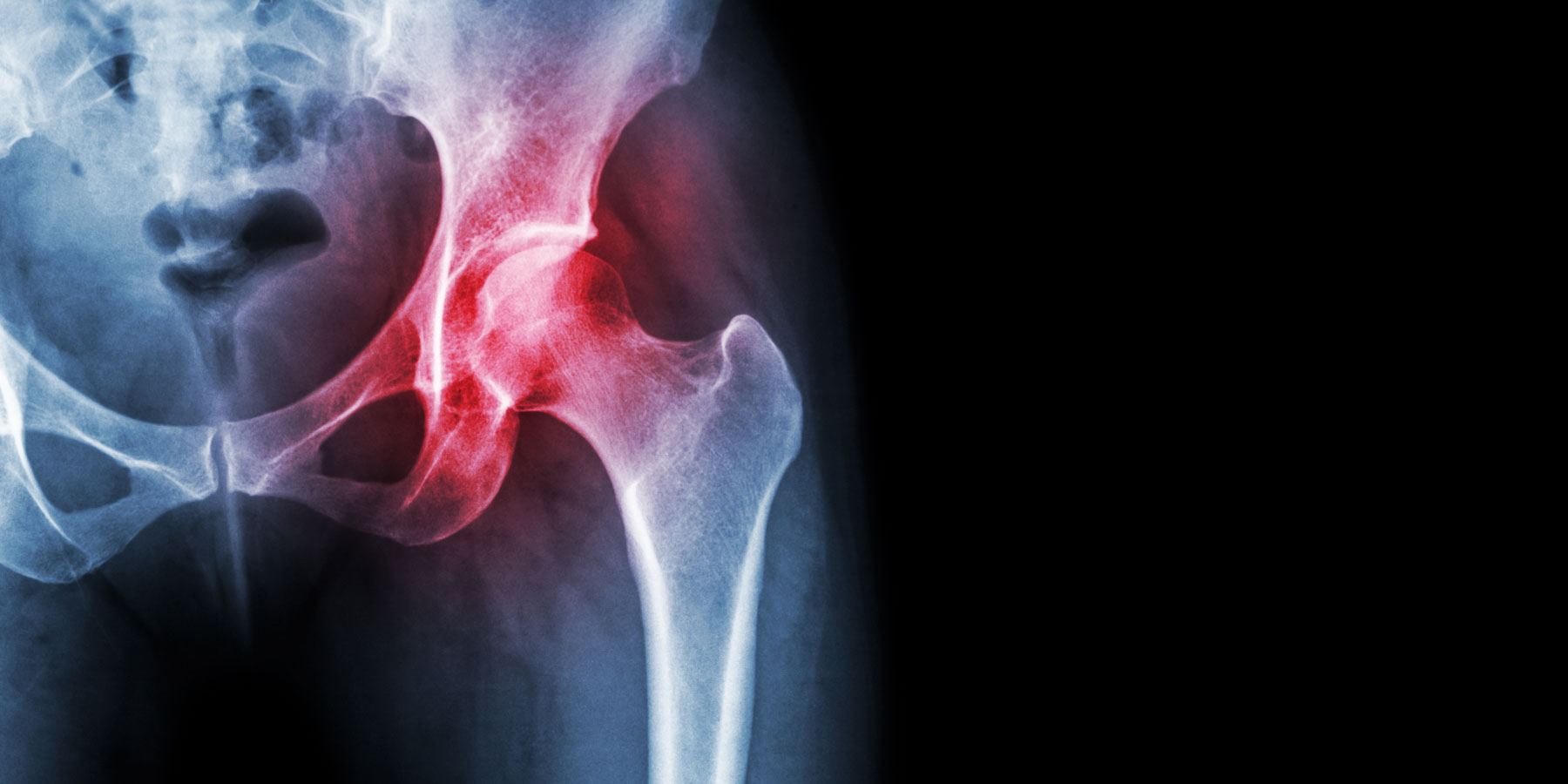
Understanding Hip Dislocations & Instability

According to the American Academy of Orthopaedic Surgeons, motor vehicle collisions are the most common cause of traumatic hip dislocations. The dislocation often occurs when the knee hits the dashboard in a collision. Wearing a seatbelt can greatly reduce your risk of hip dislocation during a collision. A fall from a significant height (such as from a ladder) or an industrial accident can also generate enough force to dislocate a hip. Hip dislocations are relatively uncommon during athletic events but can happen.

Whether you or a family member had an accident or are experiencing hip instability due to normal wear and tear, it’s smart to understand the anatomy of the hip joint, common symptoms, and the evaluation process.

Anatomy

The hip is a ball and socket joint. The femoral head (ball-shaped bone that sits at the top of the femur) and the acetabulum (socket in the pelvis that the femoral headrests) form the hip joint. The labrum is a cartilage ring that lines the acetabulum. The labrum protects the surface of the bone and provides stability. Surrounding the hip are major ligaments and muscles which offer protection and support from trauma and everyday activities.

Initial injury: Dislocation

Hip dislocation is very painful and considered an emergency due to the complications it can cause. Hip dislocation is caused when the femoral head is pushed out of the socket either backward (posterior) or forward (anterior). The most common type is posterior hip dislocation.

Hip dislocation and instability can be caused by trauma (motor vehicle accidents, sports injuries), developmental conditions (hip dysplasia) or genetic conditions. If the hip dislocation is suspected the person should not be moved and help should be called for immediately. The person will be taken to a medical facility to have the hip evaluated and relocated.

Signs of hip dislocation include:

  • Severe pain in the hip, groin, or upper leg
  • Inability to move the affected extremity
  • Shortening of the leg


After Injury

Complications from hip dislocation include fracture, hip instability, damage to the sciatic nerve, osteonecrosis (bone death), and/or arthritis. Symptoms can present immediately or take months or years to show up. The need for follow up examinations post-injury is important to rule out post-injury complications, prevent future injuries, and monitor for future conditions.

Hip instability

Damage to the ligaments surrounding the hip or to the labrum due to trauma or normal wear and tear can cause recurring hip instability. Symptoms of hip instability include:

  • Pain in the hip or groin
  • A sensation of the hip coming out of the socket
  • Hearing snapping, clicking, or popping sounds or sensations
  • Abnormal gait

If you are experiencing these symptoms it would be beneficial to have your hip evaluated to prevent future injury or dislocation. Initial evaluation of hip instability or pain would begin with a complete examination of the affected hip and x-rays would be obtained. 

At Advanced Orthopaedic Specialists we discuss the least invasive treatments first beginning with physical therapy for muscle strengthening, anti-inflammatories, and activity modification. If symptoms persist we offer further workup, injections, and surgical options. 

Here to help

If you are currently having hip pain or concerning symptoms, or you have experienced a hip injury in the past and want to prevent another one from occurring, the providers at Advanced Othopaedic Specialists are here to help. Contact us to schedule an appointment.