Understanding Dry Needling | Advanced Orthopaedic Specialists | Fayetteville, AR | Rogers, AR
Advanced Orthopaedic Specialists

February 25, 2019

| Evan Hill, DPT

Understanding Dry Needling

As physical therapists, we help our patients manage pain in a variety of ways. Dry needling is one relatively new treatment that can have great results. Dry needling involves inserting a thin, stainless steel needle directly into the involved muscle to target a myofascial trigger point, or sensitive “knot” in a muscle. The needle acts to release muscle tightness to regain normal blood flow, oxygen, and range of motion. All of which can help speed up a patient’s return to active rehabilitation.

Why Is It Called Dry Needling?  

The term “dry needling” is used because the filiform needles do not use medication or inject fluid in the involved area. Depending on the underlying issue and clinician’s discretion, the needles can be left in place for a short duration or moved in and out or “pistoned” in the muscle to release the trigger point. Because of this, other terms commonly used to describe dry needling include trigger point dry needling and intramuscular manual therapy.

It may sound intimidating, but dry needling is safe and often an effective technique used to treat a variety of sports injuries and muscle pain.

Is It Different from Acupuncture? 


Dry needling is not acupuncture, a practice based on traditional Chinese medicine and performed by acupuncturists. Both treatments use the same type of needle, but acupuncture does not target specific muscle areas. Therefore, the needle is not inserted below skin level. Acupuncture is used to target body energy flow to help relieve pain and a variety of other symptoms. Also, in contrast to dry needling, acupuncturists will apply needles in various locations on the body to achieve desired results.

What Can I Expect with Dry Needling? 


Physical therapists wear gloves, and the sterile needles are disposed of in a medical sharps collector. There are few complications to dry needling, but they do include localized bruising, bleeding, and temporary soreness.

Commonly, dry needling is used in conjunction with exercise and other treatments in a physical therapy program to achieve desired results. As with all treatments, results do vary but often patients can feel improvements with pain in as little as 1-2 treatments.

In conclusion, both dry needling and acupuncture are treatments that can provide great results but are used for different underlying impairments. It’s always advised to consult with a medical professional. 

If you’re interested in learning more about dry needling, we offer this service in our Rogers office. Give us a call or schedule an appointment to see if this is a good treatment option for you.

Written by Evan Hill, DPT
March 13, 2018

| AOSHogDocs

Traveling with the Team

One of the unique benefits of being a team physician with the University of Arkansas is accompanying the teams on certain away games. This week Dr. Yakin, Dr. Ylanan and myself travelled with the Razorback Women’s Basketball Team to Nashville, TN for the SEC Women’s Basketball tournament. Dr. Yakin (orthopedic surgeon) functions as “the bone doctor”, Dr. Ylanan (primary care sports medicine) functions as “the medicine doctor”, and I am the learner (primary care sports medicine fellow) picking up bits and pieces of sports medicine wisdom from my professors.

Yes, traveling to new cities is interesting and fun, but the role of a team physician does not go on hold during these trips. In fact more preparation takes place prior to the travel than does for home games. Anticipating the medical needs of the team prior to departure is a large part of planning. Also, being prepared for the unexpected, like the bus driver with an acute sinusitis, is an important piece of being prepared. Or being prepared for endemic illnesses that are present on international trips. Also, having anti-emetics on hand for motion sickness nausea while on long bus rides is another example of situations to be prepared for while traveling with a team.

Communication between the athletic trainers and the team physicians before a travel game takes place to prepare for any medical needs or musculoskeletal injuries that the team may have. For example, if one member of the team falls ill in the days leading up to a travel game it is our job to ensure they receive adequate prompt treatment and their teammates/roommates who may have been exposed receive necessary prophylaxis. This will ensure that the players who are healthy are able to play their game to their highest level of play.

As a physician who travels with their team you not only take care of the health needs of the athletes but also the needs of all support staff. There may not be an opportunity for someone traveling with the team to see a physician during a demanding schedule on the road with few breaks between game preparation, video, and chalk talk. From the starting point guard to Big Red, if you were requested to travel with the team you have an important role to help the team win. Often we serve as a type of concierge physician while on the road with the team.

Each provider carries their own medical bag stocked with the tools of their respective trade. Should the situation arise where someone requires medical attention the physician will have the supplies necessary to assist their patients. Ensuring this bag is prepared days prior to departure is a priority of a team physician.

Networking with the home team’s physicians and athletic trainers is not only collegial but also important for a team physician who travels. Over the years, many of the faces that you encounter while on road games are there year to year. They may not all be with the same team every year, but they tend to stay in this world. Professional friendships amongst other team physicians and athletic trainers is both give and take. If your athletes are in need of a medication for illness the home team’s physician will usually help out the visiting team’s physician with a prescription or necessary work up. The home team’s athletic trainers may also have something that your team doesn’t travel with or an extra item that your team may already be using. This in turn is reciprocated when they come to your home field or court. Coordination with the EMS and their support staff is also crucial prior to games In case there is an emergency while the game is occurring.

I have been fortunate over the last 8 months to travel as a primary care sports medicine fellow with some of the Razorback teams. I have gone to Tuscaloosa, Baton Rouge, Columbia, SC, Oxford, Dallas, Little Rock, and now Nashville with Razorback teams. There has been great experiences along the way while serving the teams on the road. “Being prepared” is probably the most important facet I have learned from my mentors while traveling with the teams but communication with home team physicians and the athletic trainers is also an integral part of being a good team physician.

December 20, 2016

| Christopher Arnold, MD

Torn ACL, Now What?

What is the Anterior Cruciate Ligament (ACL)?

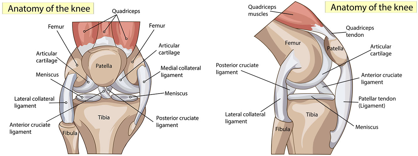
The anterior cruciate ligament (ACL) is the primary stabilizer to the knee. It offers stability to the knee during running and cutting activities. In the United States there are about 95,000 injuries per year. Most injuries occur during running, cutting, and twisting sports or activities such as soccer and basketball. The injured patient often feels a “pop” and experiences immediate swelling and inability to ambulate. Once the pain has subsided, most patients experience significant symptoms of instability with sports as well as day-to-day activities. The diagnosis is made typically by clinical examination by an orthopedic surgeon and confirmed with a MRI.

anatomy of the knee

What are my options if my ACL is torn?

Some patients who tear their ACL will elect to treat it non-operatively. They will be treated with aggressive physical therapy to strengthen the muscles around the knee. They will also be fitted for an ACL brace. They are encouraged to participate in non-impact activities, straight-line running, and avoid cutting and twisting sports. Other patients elect to have the ACL reconstructed surgically. These patients are typically younger athletes or those who participant in sports that require cutting and twisting motions.

How can the ACL be fixed?

When the ACL is torn we know it won’t heal itself. We also know that the ACL cannot be sewn back together. We do know that the current reconstruction techniques have a very high success rate. An ACL reconstruction involves placing a new graft in the place of the old ACL. The most common sources of grafts are the patellar tendon graft, the hamstring graft, and an allograft (from a cadaver). These all have high success rate. I prefer a patellar tendon graft. I harvest the graft from the same knee and through an arthroscope insert it into the place of the old ACL. The surgery typically takes one hour.

If I have my ACL fixed, what can I except afterwards?

After an ACL reconstruction the patient goes home the same day. The patient will start physical therapy immediately using a continuous passive motion machine to slowly flex the knee. The patient is able to bear weight immediately while wearing a knee brace. The knee brace is discontinued once the patient returns good strength of the knee which is typically 7 to 10 days. The patient will start to ride a stationary bicycle once the knee is able to easily bend to 90 degrees (typically two weeks). The patient is allowed to swim at approximately two months. A brace is applied at three months and the patient is allowed to run straight ahead only. At four and a half months the patients works with a trainer to being light cutting and twisting movements. A full release is given at approximately six months. We have had good success with this technique and anticipate the injured athlete will be able to resume full activities between six and twelve months.

February 21, 2017

| Christopher Arnold, MD

Too Young For a Knee Replacement

Osteoarthritis of the knee is a very common condition that affects millions of people every year. Arthritis of the knee is whenever the cartilage of the knee is lost. There are two types of cartilage. First is the meniscus. The meniscus is a rubbery cushion that comes between the two bones of the knee. It provides the initial zone of protection. Very commonly, a meniscus can be torn with day-to-day activities wear and tear, or even a high level of sports. The treatment of a meniscus tear, if it is symptomatic enough and fails to respond to therapy and/or injections, is an arthroscopic procedure. During the arthroscopy, the meniscus is either sewn back together or trimmed out, depending on the location of the tear. The second type of cartilage is articular cartilage. It is a cap that covers the top of the thigh as well as the top of the shin bone and is like gristle. Whenever damage to the articular cartilage occurs, this sets the stage for arthritis. The process goes from the cartilage cap becoming soft to small crevices, to big flaps to bare bone. Once the knee gets to bare bone, the patient experiences constant pain that can limit their activities. Typically, these patients have pain with rest as well as pain with walking short distances that is quite disabling and the end result historically has been a total knee replacement.

At Advanced Orthopedic Specialists, we established the Cartilage Restoration Center for the patients under the age of 55 who have arthritis that has progressed to the point of possibly needing a knee replacement. There are two types of cartilage restoration procedures that we perform at AOS to try to halt this progression. First is the meniscus preservation technique. If a patient has a meniscus tear which is repairable, we, through the arthroscope, can sew it back together and anticipate a good result. If; however, the patient has lost the majority of the meniscus during a prior procedure or during an injury, they could be a candidate for a meniscal allograft. The meniscal allograft is when a cadaver cartilage is taken and inserted into the knee with the assistance of an arthroscope and allowed to heal back in. We have had good success with this performing hundreds of these at Advanced Orthopedic Specialists. This is the first step to stop the progression of the arthritis.

The second type of cartilage restoration procedure involves the articular cartilage, which is the cartilage cap. There are a variety of options to restore the cartilage cap depending on the number of defects, the size of the defects, and the location of the defects. If there is a small isolated defect, the patient may be a candidate for an osteoarticular autograft (OATS). In this technique, a graft is taken from part of the knee, which is nonweightbearing and transferred to the defect in the knee. This is similar to backfilling the hole on putting green. This offers excellent results for lesions that are less than 2 sq cm.

If the lesion is larger than 2 sq cm, the options are an osteoarticular allograft, which is a large plug of cadaver cartilage, which is  transferred to the defect.

Another option is autologous chondrocyte implantation. At advanced Orthopedic Specialist, we have performed over 300 ACIs. In this technique, a knee is scoped and multiple small specimens are taken from a non-weightbearing part of the knee. They are sent to a company in Boston where they are multiplied to approximately 48 million of the patient’s own articular cells and these cells are then inserted into the knee where they are allowed to grow. This is a stem cell technique. This has had 85% success rate.

Using the combination of meniscal allografts as well as osteoarticular autografts, ACI, and steoarticular allografts, we have been able to salvage the knee in over 500 patients in the past eight years and prevent the patients under the age of 55 into having a total knee arthroplasty. We feel this is the future of knee surgery. We expect that this will ultimately replace the need for knee replacements in our patient population and the age limit of 55 will be extended.

If you have any questions about this procedure, please contact us.

May 29, 2019

| AOSHogDocs

The Difference Between Osteoporosis and Osteoarthritis

It’s National Osteoporosis month and National Osteoarthritis month, which makes this a great time to talk about these two conditions. Like many people, you may be wondering what the most significant difference is between the two. It is a very common occurrence that patients will come into our office and say they have “osteo-something.” These two terms are often confused among patients, and some think having one means they have the other. Here’s some information that may help to know.

Understanding Osteoarthritis

Osteoarthritis, commonly referred to as the “wear and tear” type of arthritis, is when the cartilage of the joint deteriorates from usage. This can affect many joints throughout the body as people use their body every day and sometimes strenuously through sports, manual labor, exercise, etc.

Everyone will develop osteoarthritis, but it may develop at different ages and be silent if it is not aggravated. Osteoarthritis is a very treatable condition if dealt with early and can be managed with physical activity, therapy, medications, injections, and cell therapy.

Understanding Osteoporosis

On the other hand, osteoporosis is when an individual has a low bone mineral density for their age. Having osteoporosis can make you more vulnerable to a fracture that can occur from a simple fall. This can be caused by certain medical conditions, genetics, poor nutrition, or may be a result of the aging process.

It is essential to make sure your dietary needs for calcium and vitamin D3 are being met. Many people take supplements, but your body absorbs these nutrients more efficiently through dairy foods or foods fortified with calcium and/or vitamin D. An individual’s maximum bone mineral density occurs somewhere between the ages of 25-35 years old, so it is important to start a healthy diet at a young age before it’s too late.

Schedule a Consult

If you feel like you have either of these conditions and would like to visit with one of our specialists about ways to manage your pain, we’d be happy to see you. Schedule an appointment with us today!

February 07, 2017

| AOSHogDocs

Stem Cell Injections

What are stem cells?

Stem cells are a biologic injection used in the sports medicine and orthopedic world to aid in injury healing. Biologics, which include stem cells or PRP (which fall under the umbrella of regenerative medicine) use growth factors (chemicals released to stimulate healing and growth) and progenitor cells (found in adipose tissue or bone marrow), to rebuild injured/damaged tissue using natures’ pathways. Stem cells contain all the genetic information allowing them the ability to mature into any cell to aid in injury healing.

How is a stem cell injection performed?

Stem cells can be obtained in using different methods. The two most common methods used are a patient’s own cells (harvested through adipose tissue aspiration or bone marrow aspiration) or through donated harvested amniotic tissue. A single stem cell injection, which is a simple in-office procedure, has the ability to stimulate healing and growth for at least 12 months. For 10 days post-procedure, certain anti-inflammatories (NSAIDs) are withheld and a targeted low-impact exercise program is started. These precautions allow the regenerative pathway to begin.

How do I know if I am a candidate for a stem cell injection?

PRP can be used for a variety of sports medicine and orthopedic injuries. PRP is currently being used in the treatment of certain tendon tears, partial rotator cuff tears, and joint arthritis. If you have a sports medicine or orthopedic injury and you are interested in seeing if stem cells are an option for you, please feel free to contact us for a consultation.

November 29, 2016

| AOSHogDocs

Skin Infections for Athletes

What is it?

There are many different types of skin conditions that can affect athletes. Causes of skin infections include viruses, bacteria, and fungi. Higher risk sports for skin infections are football and wrestling due the close skin to skin contact. However, not all infections are caused by skin to skin contact. Some may be caused by touching a contaminated surface. Athletes are at higher risk for obtaining a skin infection if they have open wounds, poor hygiene practices, or if they are sharing towels and equipment. It is important to know which type of infection an athlete has because some infections can still be passed to others after the skin is covered.

skin infections

What are the Symptoms?

It may start with the athlete noticing a new rash or lesion on his or her body. Rashes may be itchy, painful, swollen, or reddish in color. Some infections may start out looking like a bug bite or a pimple. Some athletes may also develop a fever.

When Should I See My Doctor?

Athletes should report any new skin findings to their athletic trainer, coach, or parent. If there is any concern of a skin infection the athlete should be referred to a physician. Depending on the type of infection the athlete may require topical treatment or may need to be placed on a medication. In some instances the lesion may need to be drained in the doctor’s office.

How Can I Prevent Skin Infections?

Athletes should always keep their wounds covered. It is important to shower immediately after practices or games and before using a whirlpool or other shared area. Athletes should refrain from sharing towels or razors. Uniforms should be washed and dried after each use.

When Can I Return to Play?

There are guidelines in  place by the high school federation and NCAA which state how long treatment is needed before the athlete can return to sport depending on the type of infection.  In general the athlete can return once they have been treated and the symptoms have resolved.

April 05, 2017

| AOSHogDocs

Shoulder Osteoarthritis

What is Shoulder Osteoarthritis?

Shoulder osteoarthritis (OA) is the wear and tear type of arthritis that develops over the years. It tends to be accelerated in anyone who has had trauma to their shoulder in the past. OA first starts as softening of the bone cartilage cap and then it continues to erode down to bare bone. It can be very painful at times or silent. Patients commonly describe the pain as a dull ache or a sharp pain in their shoulder. Often patients will notice a restriction in their ability to move their arm. This may make it difficult to grab the seat belt, brush your hair, or reach above your head. Often people will feel their shoulder cracking as the arthritis worsens.

How Can I Prevent Shoulder Osteoarthritis?

Shoulder osteoarthritis is very difficult to prevent, but there are certain variables that can be change to help. Individuals who do a lot of work and lifting with their arms are at a higher risk of developing osteoarthritis in their shoulder. The shoulder was not made to be a weight bearing joint, so individuals who engage in activities that place a lot of stress on their shoulder can quicken the development of osteoarthritis. If these types of activities can be decreased or avoided, then osteoarthritis can be slowed.

When Should I See My Doctor?

Shoulder osteoarthritis is progressive and it’s important to have it under control, so you can enjoy your life. Patients often seek help because the pain becomes too much, stiffness makes it difficult to move their arm, or the pain starts to wake them up at night. Ice will help control the inflammation and pain. Over the counter anti-inflammatories may also be help to again control pain and inflammation. If the pain persists despite these basic treatments, then you should see your doctor. Your physician may choose to do x-rays to check on the amount of arthritis and can often provide some treatment. Treatment may include prescription anti-inflammatories, physical therapy, or different types of injections. Injections can be used to reduce pain and inflammation or to restore cartilage and reverse arthritis. As arthritis becomes severe then the options become limited and a joint replacement may be indicated. If your shoulder has been bothering you for a prolonged period of time, then please seek the help of a sports medicine doctor.

June 07, 2017

| AOSHogDocs

Shoulder Impingement

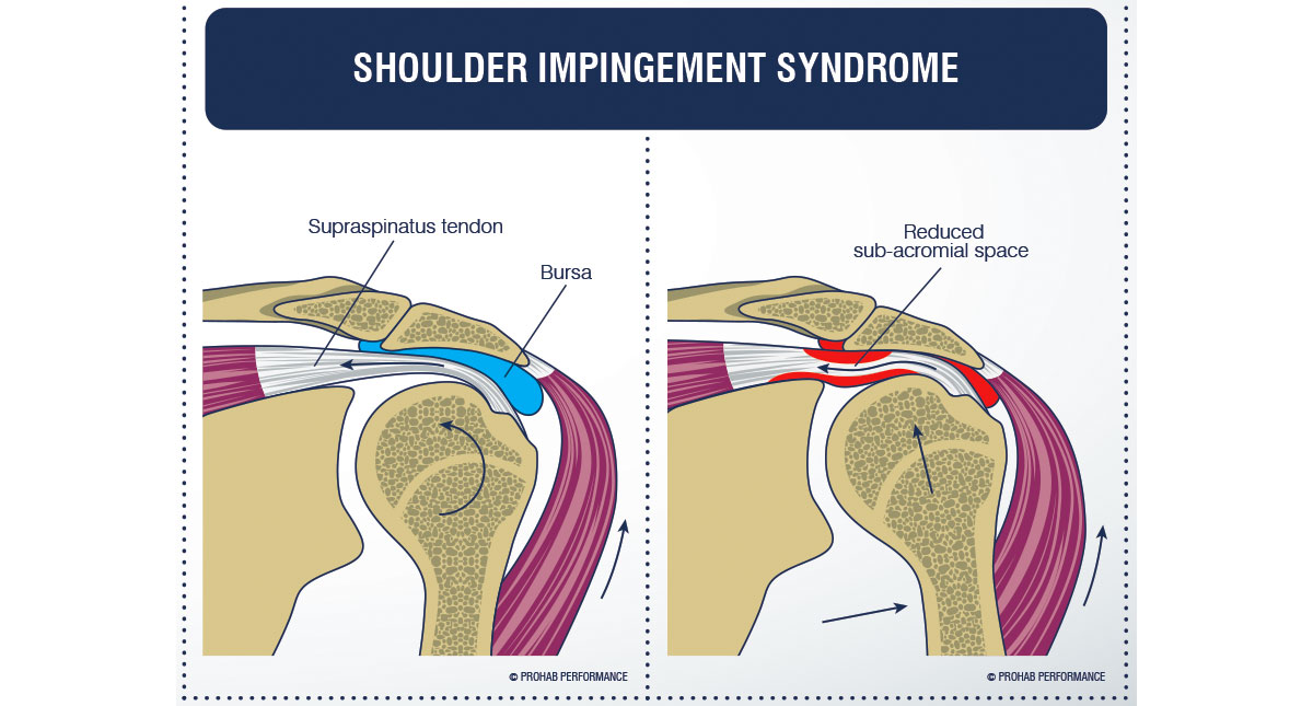
Shoulder impingement is a common condition characterized by sharp pain in the top of the shoulder when reaching or lifting your arm overhead. Often there is also pain when reaching behind the back. This pain is due to rotator cuff tendons becoming compressed or pinched as they run through the shoulder joint. If not treated, shoulder impingement can increase in severity and become very disabling.

shoulder impingement

Shoulder impingement can be caused by a variety of factors. The primary way that the tendons become compressed is when the space in the joint where the tendons run is reduced. This can be due to the development of bone spurs inside the joint space or when the top of the humerus bone is no longer aligned perfectly in the shoulder joint. Shoulder stiffness and decreased flexibility can make it difficult for the humerus to glide in the joint and decrease the space where the rotator cuff tendons run.

Muscle weakness can also lead to shoulder impingement. The rotator cuff muscles function to hold the humerus in correct alignment in the joint as the arm is raised and if they become weak or atrophied, this alignment can be lost. Injuries to the rotator cuff muscles or tears in the tendon can also lead to muscle weakness so these two conditions are commonly found together. Weakness of the shoulder blade muscles can also cause impingement when weak muscles fail to hold the shoulder blades in correct position during overhead movement.

Shoulder impingement can be successfully managed with non-surgical treatment including physical therapy. If untreated, impingement can develop into bursitis, where the tissues in the shoulder joint become irritated and inflamed. If impingement is caused by friction between bone spurs and the rotator cuff tendons, repetitive motion can damage the rotator cuff tendons which can require surgical interventions.

Surgical interventions for shoulder impingement include arthroscopic procedures to increase space in the shoulder joint including smoothing bone spurs and partially removing sections of the joint to increase space. Damaged rotator cuff tendons can also be repaired allowing for improved muscle function during overhead motion.

Sharp pain in the top of the shoulder with overhead movement or aching in the shoulder when laying on the shoulder at night should be evaluated by an orthopaedic specialist who will identify the cause of the problem and differentiate between impingement, rotator cuff injury, frozen shoulder and other disorders of the shoulder. Early identification is key to successful treatment and the prevention of surgical intervention.

January 03, 2017

| Terry Sites, MD

Rotator Cuff Injuries: Evaluation and Options

The Rotator Cuff

The rotator cuff is a group of four muscle-tendon units that originate from the shoulder blade and attach to the humeral head of the shoulder. These four muscle-tendon units (supraspinatus, infraspinatus, subscapularis and teres minor) are important in maintaining dynamic stability of the shoulder joint and to provide movement of the upper extremity away from the body such as reaching forward, backward or above shoulder level.

The supraspinatus is the most common component of the rotator cuff involved with pathology. These pathologies are one of the most common causes of shoulder pain. The pain is often felt as sharp or deep and achy, is usually located over the front corner of the shoulder and may radiate down towards the elbow. These pains are often experienced when getting dressed, reaching to the backside, reaching above shoulder level or when lying on the side. While there may be trauma associated with the onset of rotator cuff symptoms they more commonly manifest insidiously with a gradual onset. Rotator cuff pain can occur from simple inflammation of the tendon (often called tendinitis or bursitis), partial tearing or full-thickness tearing. It is often difficult clinically to separate pain generated by tendinitis from that which occurs from a complete tear. Associated symptoms may be feelings of weakness and stiffness or loss of motion. Symptoms from tendinitis often begin to present themselves in patients age 20 and beyond, rotator cuff tears generally do not occur prior to the age of 40.

Evaluation and Treatment

Physician evaluations for shoulder pain generally include a thorough history and physical examination of the shoulders and upper extremities, along with x-rays of the symptomatic shoulder. Initial treatment options include activity modification, cortisone injection, oral anti-inflammatories, and/or physical therapy. Patients who fail to improve with initial treatment modalities may be recommended to undergo an MRI examination to more fully evaluate the rotator cuff. Regular x-rays do not visualize the soft tissues of the rotator cuff, and thus an MRI can be very helpful in separating out tendinitis from rotator cuff tears in those individuals who do not improve with initial treatment.

Patients who are found to have painful rotator cuff tears have the option to undergo surgical treatment. I perform rotator cuff surgery utilizing minimally invasive techniques of arthroscopy. Surgeries are performed on an outpatient basis. Outpatient therapy is employed postop to maximize recovery and rehabilitation. Our goal is to minimize pain and maximize functional outcome.

Other conditions which can cause shoulder pain include but are not exclusively those of impingement, osteoarthritis, frozen shoulder, labral tears and biceps tendinopathy. These conditions will be addressed in future blogs, so please stay tuned.